- Home
- About
- Partners
- Newcastle University
- University of L'Aquila
- University of Manchester
- Alacris Teranostics GmbH
- University of Pavia
- Polygene
- Consiglio Nazionale delle Ricerche
- INSERM
- Certus Technology
- Charité Universitaet Medizin
- GATC Biotech
- University Medical Center Hamburg Eppendorf
- Evercyte GmbH
- University Hospital of Cologne
- PRIMM Srl
- University of Freiburg
- University of Antwerp
- Finovatis
- Research
- SYBIL at a glance
- Bone
- Growth plate
- Desbuquois dysplasia
- Diastrophic dysplasia
- MCDS
- Osteopetrosis
- Osteoporosis
- Osteogenesis imperfecta
- Prolidase deficiency
- PSACH and MED
- Systems biology
- SOPs
- Alcian Blue staining
- Bone measurements
- BrdU labelling
- Cell counting using ImageJ
- Chondrocyte extraction
- Cre genotyping protocol
- DMMB assay for sulphated proteoglycans
- Densitometry using ImageJ
- Double immunofluorescence
- Electron microscopy of cartilage - sample prep
- Extracting DNA for genotyping
- Grip strength measurement
- Histomorphometry on unon-decalcified bone samples
- Immunocytochemistry
- Immunofluorescence
- Immunohistochemistry
- Quantitative X-ray imaging on bones using Faxitron and ImageJ
- Skeletal preps
- TUNEL assay (Dead End Fluorimetric Kit, Promega)
- Toluidine Blue staining
- Toluidine Blue staining
- Von Kossa Gieson staining
- Wax embedding of cartilage tissue
- Contact Us
- News & Events
- Links
- Portal
Growth plate
The growth plate or physis is the structure responsible for longitudinal bone growth and endochondral ossification. The growth plates in humans first appear at the end of the first trimester of pregnancy and fuse when the growth terminates. The growth plate consists of several distinct zones, reflecting the gradual transition of cells through different stages of differentiation. Cells at the same level in the growth plate are at exactly the same stage of development. Chondrocytes in the physis remain spatially fixed all their life in a so called “lacunae”, but are able to modify their environment and interact with it by changes in protein expression and in this way influence the matrix in which they reside.
The distinct zones found in the cartilage growth plate are:
- resting zone
- proliferating zone
- hypertrophic zone
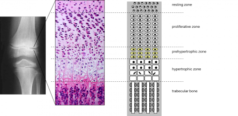
The resting zone is located at the top of the growth plate. It acts as a reserve of precursor cells for the proliferating chondrocytes in the columns, where cells reside prior to entering the accelerated proliferation stage, embedded in rich extracellular matrix. The cells of the resting zone are round and have a high lipid content suggesting a nutrient storage potential.
The proliferating zone comprises at least half of the overall height of the physis, depending on the age. In this zone the cells flatten and divide, forming characteristic columns and laying down a cartilage extracellular matrix that will later serve as a scaffold for bone formation.
In the hypertrophic zone the chondrocytes are still organised in columns, but the cells and their lacunae become 5-12 times bigger. The amount of extracellular matrix also increases 3 fold, and the matrix composition is altered, with specific chondrocyte hypertrophy markers such as type X collagen and alkaline phosphatase present. The cells eventually die, triggering blood vessel invasion and bone formation.
Bone formation in the process of endochondral ossification depends on blood vessel invasion and matrix remodelling. At the last stages of the process the cartilage extracellular matrix is degraded by hypertrophic chondrocytes themselves and by chondroclasts and preosteoclasts brought in by newly forming blood vessels. The blood vessels then enter the partially dismantled scaffold, bringing osteogenic progenitor cells that in turn deposit trabecular bone. Several growth factors and proteolytic enzymes are implicated in this process. The remodelling processes cause variations in calcium and phosphate ions in the matrix at the junction between cartilage and bone. The variations in ion concentrations, matrix degradation and mineralisation, and influx of new cell types all regulate the equilibrium of cartilage degradation to bone formation.